Adding Tension to the Sacrotuberous and Sacrospinous Ligaments
The sacrotuberous ligament connects the sitting bones to the sacrum.
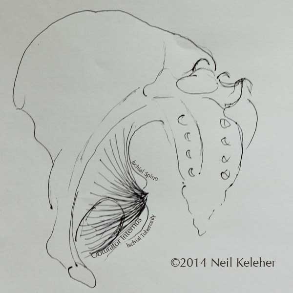
The sacrospinous ligament attaches from the ischial spine (just above the ischial tuberosities, and in between the upper and lower sciatic notches) to the sacrum.
Pulling the sitting bones apart, and nodding the sacrum forwards so that the tailbone is flicked backwards (nutation) can add tension to these ligaments.
Muscles that Attach to the Sacrotuberous Ligament
Two muscles that attach to the sacrotuberous ligament are the gluteus maximus and the biceps femoris. (The piriformis also has attachments to this ligament.)
Gluteus Maximus Attaches to the Sacrotuberous Ligament
The gluteus maximus has fibers that attach from the sacral tuberous ligament to the thigh. Adding tension to this ligament, by spreading the sitting bones, potentially gives these fibers of the gluteus maximus a more solid foundation from which to act.
The Hamstrings Connect to the Sacrotuberous Ligament.
The hamstrings connect to the sitting bones and so potentially, adding tension to the sacrotuberous ligament can create a line of tension all the way from below the knees to the sacrum, and potentially from there up the back of the spine.
And so spreading the sitting bones using the internal obturators can be a key action in forward bends since it integrates the back line of the body.
The biceps femoris long head in particular has fibers that are continuous with those of the sacrotuberous ligament.
Now what's so important about the obturator internus?
Obturator Internus Has A Large Area of Attachment Within the Pelvis
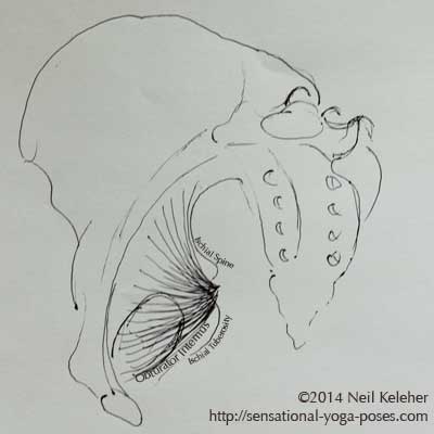
The obturator internus has a large area of attachment within the bottom back half of the pelvis.
If you draw a line from the top rear of the pelvis (just above the upper surface of the sacrum) to the pubic bone, the obturator internus attaches to most of the area below this line.
In part, it attaches to connective tissue covering the obturator foramen, the openings at either side of the bottom of the pelvis. This is what gives it its name.
(The obturator externus attaches to the outside of the connective tissue covering this opening.)
The Obturator Wraps around the Back of the Hip Bone
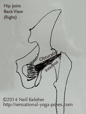
The obturator internus wraps around the lower sciatic notch, which is just above the sitting bone. From there, assuming a standing upright position, it passes forwards and upwards to attach near the 12 o'clock position to the inner surface of the greater trochanter, near where it connects to the neck of the thigh.
Acting from thighs that are stabilized against external rotation, the obturator internus can be used to pull the sitting bones apart.
It can also be used to exert a forward pull on the hip bone, relative to the femur.
The Gemelli Twins
After passing around the lower sciatic notch the obturator internus unites with the two gemelli muscles which attach to the ischium just above and below the sciatic notch.
These attach to the same point on the greater trochanter as the obturator internus.
These muscles can work together in spreading the sitting bones.
Controlling the Shape of the Pelvis
I believe that the large surface area to which obturator internus attaches gives it a broad foundation from which to act, and so perhaps a greater ability to act in spreading the sitting bones.
This actually results in a shape change of the pelvis, slight but perceptible. This shape change can affect tension in the pelvic floor as well as the hips joints as well as the lumbar spine.
Tilting the Sacrum Rearwards (Counter-Nutation)
The pelvic floor muscles are one set of muscles that can be used to draw the sitting bones inwards and the tailbone forwards, towards the pubic bone, reducing the diameter of the bottom opening of the pelvis.
This same action causes the sacrum to nod backwards at the SI joint (counter-nutation).
With the two halves of the pelvis pivoting at the SI Joints and the pubic bone, when the sitting bones move inwards, (and the bottom of the sacrum forwards) the upper front parts of the pelvis (the ASICs or "eye bones" of the pelvis) spread apart and the top of the sacrum moves backwards. This causes the diameter of the top opening of the pelvis to increase.
Nodding the Sacrum Forwards (Nutation)
Nodding the sacrum forwards has the opposite effect, increasing the diameter of the bottom opening of the pelvis and reducing the diameter of the top opening. Top opening diameter is decreased by the top of the sacrum moving forwards while the ASICs move inwards. At the same time, bottom opening diameter is increased by the bottom tip of the sacrum moving rearwards and the ischial tuberosities moving outwards.
This can be driven by the transverse abdominis. But it can also be caused by the obturator internus.
Why might the pelvis and SI Joints be built with this ability to nutate and counter-nutate?
Shaping the Pelvis for Room-to-Move and Optimum Hip Joint Tension
The shoulder blades move relatively freely with respect to the ribcage. They can be positioned so that the shoulder joint always has room to move and so that the muscles that cross the shoulder joint are at optimum length for optimum tension.
As an example, when lifting the arm bones the bottom tips of the shoulder blades rotate outwards so that the arm bone can clear the accromion process, the shelf of bone that sits over each shoulder joint.
Faulty shoulder blade positioning can lead to shoulder impingement or shoulder pain or shoulders that operate poorly.
The pelvis is a more stable structure but, it may change shape to allow optimum hip muscle tension and positioning of each half of the pelvis relative to the thigh bones.
- When moving the thigh bones forwards or outwards it may be desirable to make the front of the pelvis narrower.
- When moving the thigh bones rearwards it may be desirable to make the bottom rear of the pelvis narrower.
Where pulling the sitting bones together can be helpful in backbending the hips, spreading the sitting bones can be helpful in postures where the hips are bent forwards.
In side-to-side splits, depending on the tilt of your pelvis relative to your thigh bones, either option may be appropriate.
The Obturator Internus and External Rotation of the Hip Joint
The obturator internus can act as an external rotator.
In a standing position, the natural tendency is for the arches of the feet to collapse rolling the shins and thighs inwards.
(If while standing you relax your arches, and then relax your hips, while staying upright, you may be able to experience this for yourself.)
- The muscles of the foot counter act this tendency by lifting the center of the inner arch and causing the shins to roll outwards relative to the feet.
- At the top end of the leg, the obturator internus may counter act this tendency by acting to prevent the thigh from collapsing inwards.
With both the hip and the foot working together, torsional stresses on the knee may be reduced.
Protecting An Injury to the Inner Knee (Inside of the Knee)
I had a knee injury from a motorcycle accident about twelve years ago. I never had it checked but the inner knee was damaged, to the point where it would dislocated slightly when I was walking.
Later on the knee did get better but then I found I had problems balancing on that one foot.
I believe that one of the ways that my body may have compensated for the injury, or acted to protect my knee, was to keep the obturator internus activated so that my thigh wouldn't roll inwards.
Rolling inwards perhaps places the most stress on the inner knee, and so when damaged, the body acts to protect the damaged area.
My own experience was that I couldn't ground through the base of my big toe while trying to balance on that foot
My hip isn't a hundred percent now. As well as injuring the knee I've also bruised my tailbone but it is getting better and I think the obturator internus is one of the key components in standing on one leg and in some types of seated forward bends.
One of the things that I notice now is that I tend to sit with my left sitting bone slightly lifted. The left knee was the knee I injured. This is accompanied by a slight tension in my pelvis around the region of my left sitting bone.
Conscious of it I am getting better at relaxing that tension and sitting evenly on both sitting bones.
Balancing External Rotation with Internal Rotation
As mentioned, the obturator internus may be useful in spreading the sitting bones.
This adds tension to the sacrotuberous ligament so that fibers of the glute max that attach to that ligament can also be used. And it creates a continuous line of tension from hamstrings to sacrum to the spinal erectors.
It also can cause the front of the pelvis at the ASICs to narrow even as it tries to rotate the femurs outwards.
The narrowing of the ASICs may create tension in the tensor fascae latae muscles, or give them room to contract. It may affect the gluteus minimus in the same way.
Gluteus Minimus and Tensor Fascia Latae
Both muscles attach at or near the ASICs. Both can help cause internal rotation (whereas the obturator internus tends to cause external rotation).
Tensor fascae latae (TFL) attaches to the IT Band which runs down the outside of the thigh, over the vastus lateralis muscle. It attaches to the top of the tibia, just in front of the fibula.
It can help to internally rotate the shin and thigh together if the knee is straight.
Gluteus minimis attaches to the outer surface of the pelvis, roughly between the ASIC and the hip socket. It can help to rotate the thigh internally.
With TFL and/or glute minimus active, the external rotation from the obturator internus can be nullified or balanced.
One Legged Standing Forward Bend With the Lifted Knee Pulled Forwards
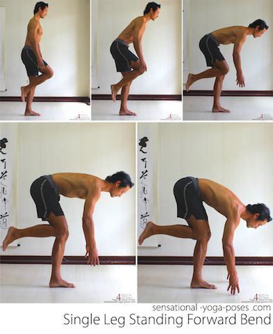
One of my favorite exercises for stabilizing the hip is the one legged standing forward bend.
The key difference between this and a pose like warrior 3 is that the knee of the free leg is bent and pulled forwards and kept pulled forwards.
You could also do it with the lifted knee straight as described here in hip stability exercises
In either case, with the lifted knee or leg pulled forwards, the hips have to shift back to keep your center of gravity over your supporting foot. The are stretched while being active and both they and the butt muscles have to be strong to return your to standing.
This means that the back line of the leg has to be integrated. And based on my experience so far that means activating the obturator internus to add tension to the sacro-tuberous ligament.
Spreading the Sitting Bone While On One Leg
Spreading the sitting bones is easy when both feet are on the floor so that the legs are stabile.
How do you spread the sitting bones while standing on one leg?
It's a little like flexing the biceps. (You keep the arm bones still while trying to close the front of the elbow joint.)
To activate the obturator internus while standing on one leg, you keep the pelvis stationary relative to the standing leg while "spreading" the sitting bone of that leg.
That means keeping the pelvis stable relative to the leg while spreading the sitting bone.
While tilting the pelvis forwards, limit the amount that the pelvis turns left or right so that the obturator internus can continue to stay active.
If I can't spread the sitting bone, then I'll adjust the position of my pelvis, often turning it to the lifted leg side so that I increase the space between the back of the thigh and the back of the pelvis.
This is a very slight movement.
From there I try to keep obturator internus active as I bend forwards and as I stand up.
This same feeling can then be applied in standing forward bends where both feet are on the floor.
Strong Pulling Sensations Or Sharp Tension is an Indicator of
Less than Ideal Relationship Between Pelvis and Thigh Bone
On my less strong side I find that I get a pulling sensation (uncomfortable) which means that my hip isn't working properly. And so I am now working on maintaining the ideal alignment of my pelvis while bending forwards so that my obturator internus can activate.
Ideal alignment is something that I focus on feeling so that I don't have to think about it.
If there is pain then I haven't got ideal alignment. If bending forwards and standing up is pain free, or sharp tension free, then I have got ideal alignment, or perhaps not ideal, but good enough that my body can function effectively.
Ideal, or "functional" alignment means positioning the pelvis relative to the thigh bone so that hip muscles like the obturator internus have room to activate.
If the hip muscles as a whole have room to activate then they can work effectively together to maintain the position of the pelvis or change it with a minimum of effort or sharp pain.
Published: 2011 10 03
Updated: 2020 08 04
